SWISS-MODEL Homology Modelling Report |
Model Building Report
This document lists the results for the homology modelling project "R2PDM0-MurB-E-faecium" submitted to SWISS-MODEL workspace on Oct. 17, 2017, 3:46 p.m..The submitted primary amino acid sequence is given in Table T1.
If you use any results in your research, please cite the relevant publications:
Marco Biasini; Stefan Bienert; Andrew Waterhouse; Konstantin Arnold; Gabriel Studer; Tobias Schmidt; Florian Kiefer; Tiziano Gallo Cassarino; Martino Bertoni; Lorenza Bordoli; Torsten Schwede. (2014). SWISS-MODEL: modelling protein tertiary and quaternary structure using evolutionary information. Nucleic Acids Research (1 July 2014) 42 (W1): W252-W258; doi: 10.1093/nar/gku340.Arnold, K., Bordoli, L., Kopp, J. and Schwede, T. (2006) The SWISS-MODEL workspace: a web-based environment for protein structure homology modelling. Bioinformatics, 22, 195-201.
Benkert, P., Biasini, M. and Schwede, T. (2011) Toward the estimation of the absolute quality of individual protein structure models. Bioinformatics, 27, 343-350
Results
The SWISS-MODEL template library (SMTL version 2017-10-11, PDB release 2017-10-06) was searched with Blast (Altschul et al., 1997) and HHBlits (Remmert, et al., 2011) for evolutionary related structures matching the target sequence in Table T1. For details on the template search, see Materials and Methods. Overall 164 templates were found (Table T2).
Models
The following models were built (see Materials and Methods "Model Building"):
Model #01 | File | Built with | Oligo-State | Ligands | GMQE | QMEAN |
|---|---|---|---|---|---|---|
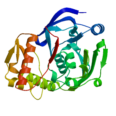 | PDB | ProMod3 Version 1.0.2. | MONOMER | None | 0.77 | -0.54 |
|  |  |
| Template | Seq Identity | Oligo-state | Found by | Method | Resolution | Seq Similarity | Range | Coverage | Description |
|---|---|---|---|---|---|---|---|---|---|
| 3tx1.1.A | 56.94 | monomer | BLAST | X-ray | 2.69Å | 0.46 | 13 - 300 | 0.93 | UDP-N-acetylenolpyruvoylglucosamine reductase |
| Ligand | Added to Model | Description |
|---|---|---|
| FAD | ✕ - Binding site not conserved. | FLAVIN-ADENINE DINUCLEOTIDE |
| GOL | ✕ - Not biologically relevant. | GLYCEROL |
| GOL | ✕ - Not biologically relevant. | GLYCEROL |
| SO4 | ✕ - Not biologically relevant. | SULFATE ION |
| SO4 | ✕ - Not biologically relevant. | SULFATE ION |
Target    MNKEEIVESLATITLLFDEPLMNYTFTKTGGPADVLAFPKKQEEVKQIIDYCRIHDISWMVLGNASNLIVQDGGIRGVVI
3tx1.1.A  ------------IAIKLNEPLSKYTYTKTGGAADVFVMPKTIEEAQEVVAYCHQNKIPLTILGNGSNLIIKDGGIRGVIL

Target    MLTEMKQIHVKGTMVIAEAGASLIDTTYAALAESLTGFEFACGIPGSVGGAVYMNAGAYGGEIKDVFAEVDLLLEDGTLK
3tx1.1.A  HLDLLQTIERNNTQIVAMSGAKLIDTAKFALNESLSGLEFACGIPGSIGGALHMNAGAYGGEISDVLEAATVLTQTGELK

Target    TLTKEEMVFSYRHSKVQELRAIVLEARFSLQTGDYEAIKARMDELTELRQSKQPLEYPSCGSVFKRPVGHYTGQLIQQAG
3tx1.1.A  KLKRSELKAAYRFSTIAEKNYIVLDATFSLALEEKNLIQAKMDELTAAREAKQPLEYPSCGSVFKRPPGHFAGKLIQDSG

Target    LQGLKWGGAQISEKHAGFIVNIDHATATDYIELIAHIQEVIKEKFDVSLETEVRIIGEKQALSETSRSLAN
3tx1.1.A  LQGHIIGGAQVSLKHAGFIVNIGGATATDYMNLIAYVQQTVREKFDVELETEVKIIGEDK-----------
Model #02 | File | Built with | Oligo-State | Ligands | GMQE | QMEAN |
|---|---|---|---|---|---|---|
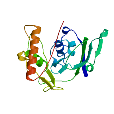 | PDB | ProMod3 Version 1.0.2. | MONOMER | None | 0.46 | -4.21 |
|  |  |
| Template | Seq Identity | Oligo-state | Found by | Method | Resolution | Seq Similarity | Range | Coverage | Description |
|---|---|---|---|---|---|---|---|---|---|
| 3i99.1.A | 37.50 | monomer | BLAST | X-ray | 2.20Å | 0.37 | 84 - 298 | 0.69 | UDP-N-acetylenolpyruvoylglucosamine reductase |
| Ligand | Added to Model | Description |
|---|---|---|
| FAD | ✕ - Binding site not conserved. | FLAVIN-ADENINE DINUCLEOTIDE |
| PO4 | ✕ - Not biologically relevant. | PHOSPHATE ION |
| PO4 | ✕ - Not biologically relevant. | PHOSPHATE ION |
Target    MNKEEIVESLATITLLFDEPLMNYTFTKTGGPADVLAFPKKQEEVKQIIDYCRIHDISWMVLGNASNLIVQDGGIRGVVI
3i99.1.A  --------------------------------------------------------------------------------

Target    MLTEMKQIHVKGTMVIAEAGASLIDTTYAALAESLTGFEFACGIPGSVGGAVYMNAGAYGGEIKDVFAEVD-LLLEDGTL
3i99.1.A  ---DYHRLHVAG----GEDWPSLVSW---CVEQGIGGLENLALIPGCAGSAPIQNIGAYGVEFKDVCDYVEYLCLETGTV

Target    KTLTKEEMVFSYR-----HSKVQELRAIVLEARFS------LQTGDYE------AIKARMDELTELRQSK--QPLEYPSC
3i99.1.A  KRLTMEECQFGYRDSIFKHQLYQKAVVTAVGLKFAKAWQPIIQYGPLKDLSSDCAIHDVYQRVCATRMEKLPDPAVMGNA

Target    GSVFKRPV-------------GHYTGQLIQQAGLQGLKW------------GGAQISEKHAGFIVNIDHATATDYIELIA
3i99.1.A  GSFFKNPVISQQAFARLQIEHPDVVAYPAEQGVKVAAGWLIDQAGLKGHQIGGAKVHPKQALVIVNTGDASAQDVLMLAA

Target    HIQEVIKEKFDVSLETEVRIIGE------KQALSETSRSLAN
3i99.1.A  DIQQRVFNCYGIELEHEVRFIGESEETNLKQWMSEQA-----
Materials and Methods
Template Search
Template search with Blast and HHBlits has been performed against the SWISS-MODEL template library (SMTL, last update: 2017-10-11, last included PDB release: 2017-10-06).
The target sequence was searched with BLAST (Altschul et al., 1997) against the primary amino acid sequence contained in the SMTL. A total of 15 templates were found.
An initial HHblits profile has been built using the procedure outlined in (Remmert, et al., 2011), followed by 1 iteration of HHblits against NR20. The obtained profile has then be searched against all profiles of the SMTL. A total of 149 templates were found.
Template Selection
For each identified template, the template's quality has been predicted from features of the target-template alignment. The templates with the highest quality have then been selected for model building.
Model Building
Models are built based on the target-template alignment using ProMod3. Coordinates which are conserved between the target and the template are copied from the template to the model. Insertions and deletions are remodelled using a fragment library. Side chains are then rebuilt. Finally, the geometry of the resulting model is regularized by using a force field. In case loop modelling with ProMod3 fails, an alternative model is built with PROMOD-II (Guex, et al., 1997).
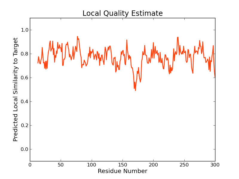
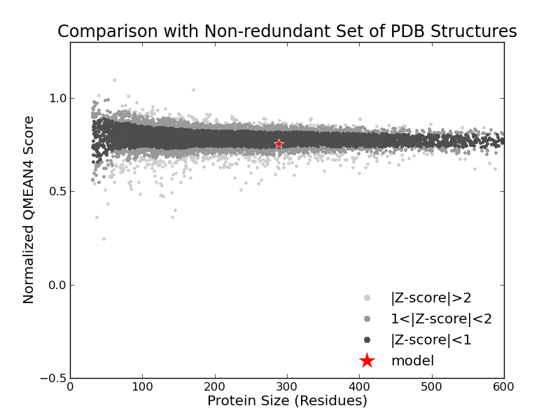
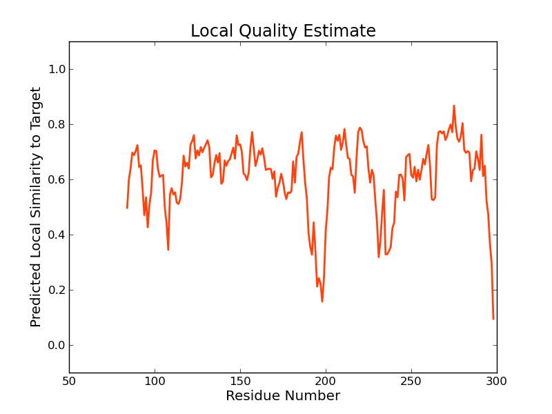
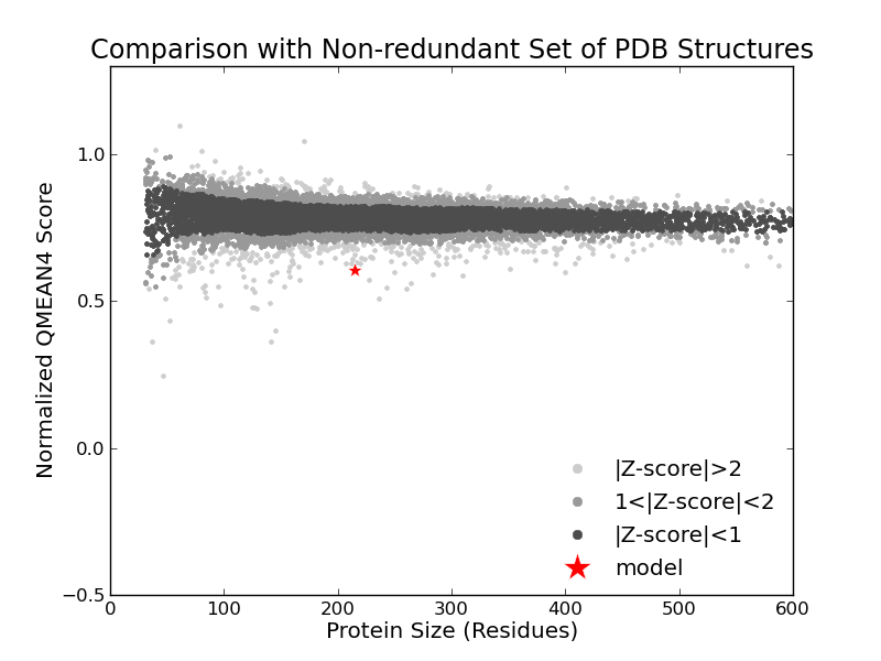
Model Quality Estimation
The global and per-residue model quality has been assessed using the QMEAN scoring function (Benkert, et al., 2011) . For improved performance, weights of the individual QMEAN terms have been trained specifically for SWISS-MODEL.
Ligand Modelling
Ligands present in the template structure are transferred by homology to the model when the following criteria are met (Gallo -Casserino, to be published): (a) The ligands are annotated as biologically relevant in the template library, (b) the ligand is in contact with the model, (c) the ligand is not clashing with the protein, (d) the residues in contact with the ligand are conserved between the target and the template. If any of these four criteria is not satisfied, a certain ligand will not be included in the model. The model summary includes information on why and which ligand has not been included.
Oligomeric State Conservation
Homo-oligomeric structure of the target protein is predicted based on the analysis of pairwise interfaces of the identified template structures. For each relevant interface between polypeptide chains (interfaces with more than 10 residue-residue interactions), the QscoreOligomer (Mariani et al., 2011) is predicted from features such as similarity to target and frequency of observing this interface in the identified templates (Kiefer, Bertoni, Biasini, to be published). The prediction is performed with a random forest regressor using these features as input parameters to predict the probability of conservation for each interface. The QscoreOligomer of the whole complex is then calculated as the weight-averaged QscoreOligomer of the interfaces. The oligomeric state of the target is predicted to be the same as in the template when QscoreOligomer is predicted to be higher or equal to 0.5.
References
Altschul, S.F., Madden, T.L., Schaffer, A.A., Zhang, J., Zhang, Z., Miller, W. and Lipman, D.J. (1997) Gapped BLAST and PSI-BLAST: a new generation of protein database search programs. Nucleic Acids Res, 25, 3389-3402.
Remmert, M., Biegert, A., Hauser, A. and Soding, J. (2012) HHblits: lightning-fast iterative protein sequence searching by HMM-HMM alignment. Nat Methods, 9, 173-175.
Guex, N. and Peitsch, M.C. (1997) SWISS-MODEL and the Swiss-PdbViewer: an environment for comparative protein modeling. Electrophoresis, 18, 2714-2723.
Sali, A. and Blundell, T.L. (1993) Comparative protein modelling by satisfaction of spatial restraints. J Mol Biol, 234, 779-815.
Benkert, P., Biasini, M. and Schwede, T. (2011) Toward the estimation of the absolute quality of individual protein structure models. Bioinformatics, 27, 343-350.
Mariani, V., Kiefer, F., Schmidt, T., Haas, J. and Schwede, T. (2011) Assessment of template based protein structure predictions in CASP9. Proteins, 79 Suppl 10, 37-58.
Table T1:
Primary amino acid sequence for which templates were searched and models were built.
ASLIDTTYAALAESLTGFEFACGIPGSVGGAVYMNAGAYGGEIKDVFAEVDLLLEDGTLKTLTKEEMVFSYRHSKVQELRAIVLEARFSLQTGDYEAIKA
RMDELTELRQSKQPLEYPSCGSVFKRPVGHYTGQLIQQAGLQGLKWGGAQISEKHAGFIVNIDHATATDYIELIAHIQEVIKEKFDVSLETEVRIIGEKQ
ALSETSRSLAN
Table T2:
| Template | Seq Identity | Oligo-state | Found by | Method | Resolution | Seq Similarity | Coverage | Description |
|---|---|---|---|---|---|---|---|---|
| 3tx1.1.A | 55.59 | monomer | HHblits | X-ray | 2.69Å | 0.45 | 0.95 | UDP-N-acetylenolpyruvoylglucosamine reductase |
| 1hsk.1.A | 51.17 | monomer | HHblits | X-ray | 2.30Å | 0.44 | 0.96 | UDP-N-ACETYLENOLPYRUVOYLGLUCOSAMINE REDUCTASE |
| 3tx1.1.A | 56.94 | monomer | BLAST | X-ray | 2.69Å | 0.46 | 0.93 | UDP-N-acetylenolpyruvoylglucosamine reductase |
| 1hsk.1.A | 54.09 | monomer | BLAST | X-ray | 2.30Å | 0.45 | 0.90 | UDP-N-ACETYLENOLPYRUVOYLGLUCOSAMINE REDUCTASE |
| 4pyt.1.A | 44.90 | monomer | BLAST | X-ray | 1.85Å | 0.42 | 0.95 | UDP-N-acetylenolpyruvoylglucosamine reductase |
| 4pyt.1.A | 45.30 | monomer | HHblits | X-ray | 1.85Å | 0.42 | 0.92 | UDP-N-acetylenolpyruvoylglucosamine reductase |
| 5jzx.1.A | 28.87 | homo-dimer | HHblits | X-ray | 2.20Å | 0.33 | 0.94 | UDP-N-acetylenolpyruvoylglucosamine reductase |
| 5jzx.1.B | 28.87 | homo-dimer | HHblits | X-ray | 2.20Å | 0.33 | 0.94 | UDP-N-acetylenolpyruvoylglucosamine reductase |
| 3i99.1.A | 29.72 | monomer | HHblits | X-ray | 2.20Å | 0.34 | 0.92 | UDP-N-acetylenolpyruvoylglucosamine reductase |
| 1mbb.1.A | 28.62 | monomer | HHblits | X-ray | 2.30Å | 0.34 | 0.91 | URIDINE DIPHOSPHO-N-ACETYLENOLPYRUVYLGLUCOSAMINE REDUCTASE |
| 1mbt.1.A | 28.62 | monomer | HHblits | X-ray | 3.00Å | 0.34 | 0.91 | URIDINE DIPHOSPHO-N-ACETYLENOLPYRUVYLGLUCOSAMINE REDUCTASE |
| 2q85.1.A | 28.62 | monomer | HHblits | X-ray | 2.51Å | 0.34 | 0.91 | UDP-N-acetylenolpyruvoylglucosamine reductase |
| 4jay.1.A | 29.08 | monomer | HHblits | X-ray | 2.23Å | 0.34 | 0.91 | UDP-N-acetylenolpyruvoylglucosamine reductase |
| 4jb1.1.A | 29.08 | monomer | HHblits | X-ray | 2.10Å | 0.34 | 0.91 | UDP-N-acetylenolpyruvoylglucosamine reductase |
| 1uxy.1.A | 28.37 | monomer | HHblits | X-ray | 1.80Å | 0.34 | 0.91 | URIDINE DIPHOSPHO-N-ACETYLENOLPYRUVYLGLUCOSAMINE REDUCTASE |
| 2mbr.1.A | 28.83 | monomer | HHblits | X-ray | 1.80Å | 0.34 | 0.90 | URIDINE DIPHOSPHO-N-ACETYLENOLPYRUVYLGLUCOSAMINE REDUCTASE |
| 2gqu.1.A | 38.40 | monomer | HHblits | X-ray | 1.60Å | 0.37 | 0.85 | UDP-N-Acetylenolpyruvylglucosamine Reductase |
| 2gqt.1.A | 38.40 | monomer | HHblits | X-ray | 1.30Å | 0.37 | 0.85 | UDP-N-Acetylenolpyruvylglucosamine Reductase |
| 4jay.1.A | 30.38 | monomer | BLAST | X-ray | 2.23Å | 0.34 | 0.84 | UDP-N-acetylenolpyruvoylglucosamine reductase |
| 4jb1.1.A | 30.38 | monomer | BLAST | X-ray | 2.10Å | 0.34 | 0.84 | UDP-N-acetylenolpyruvoylglucosamine reductase |
| 2gqu.1.A | 40.16 | monomer | BLAST | X-ray | 1.60Å | 0.38 | 0.78 | UDP-N-Acetylenolpyruvylglucosamine Reductase |
| 2gqt.1.A | 40.16 | monomer | BLAST | X-ray | 1.30Å | 0.38 | 0.78 | UDP-N-Acetylenolpyruvylglucosamine Reductase |
| 1mbb.1.A | 34.89 | monomer | BLAST | X-ray | 2.30Å | 0.36 | 0.76 | URIDINE DIPHOSPHO-N-ACETYLENOLPYRUVYLGLUCOSAMINE REDUCTASE |
| 1mbt.1.A | 34.89 | monomer | BLAST | X-ray | 3.00Å | 0.36 | 0.76 | URIDINE DIPHOSPHO-N-ACETYLENOLPYRUVYLGLUCOSAMINE REDUCTASE |
| 2q85.1.A | 34.89 | monomer | BLAST | X-ray | 2.51Å | 0.36 | 0.76 | UDP-N-acetylenolpyruvoylglucosamine reductase |
| 2mbr.1.A | 34.89 | monomer | BLAST | X-ray | 1.80Å | 0.36 | 0.76 | URIDINE DIPHOSPHO-N-ACETYLENOLPYRUVYLGLUCOSAMINE REDUCTASE |
| 1uxy.1.A | 34.47 | monomer | BLAST | X-ray | 1.80Å | 0.36 | 0.76 | URIDINE DIPHOSPHO-N-ACETYLENOLPYRUVYLGLUCOSAMINE REDUCTASE |
| 5jzx.1.A | 32.07 | homo-dimer | BLAST | X-ray | 2.20Å | 0.35 | 0.76 | UDP-N-acetylenolpyruvoylglucosamine reductase |
| 5jzx.1.B | 32.07 | homo-dimer | BLAST | X-ray | 2.20Å | 0.35 | 0.76 | UDP-N-acetylenolpyruvoylglucosamine reductase |
| 3i99.1.A | 37.50 | monomer | BLAST | X-ray | 2.20Å | 0.37 | 0.69 | UDP-N-acetylenolpyruvoylglucosamine reductase |
| 2uuu.1.A | 15.68 | homo-dimer | HHblits | X-ray | 1.95Å | 0.29 | 0.59 | ALKYLDIHYDROXYACETONEPHOSPHATE SYNTHASE |
| 2uuv.2.B | 15.68 | homo-dimer | HHblits | X-ray | 1.99Å | 0.29 | 0.59 | ALKYLDIHYDROXYACETONEPHOSPHATE SYNTHASE |
| 4bc7.1.A | 13.81 | homo-dimer | HHblits | X-ray | 2.40Å | 0.28 | 0.58 | ALKYLDIHYDROXYACETONEPHOSPHATE SYNTHASE, PEROXISOMAL |
| 4bby.1.A | 13.81 | homo-dimer | HHblits | X-ray | 1.90Å | 0.28 | 0.58 | ALKYLDIHYDROXYACETONEPHOSPHATE SYNTHASE, PEROXISOMAL |
| 4bca.1.A | 13.89 | homo-dimer | HHblits | X-ray | 2.40Å | 0.28 | 0.58 | ALKYLDIHYDROXYACETONEPHOSPHATE SYNTHASE, PEROXISOMAL |
| 3pm9.1.A | 21.59 | homo-dimer | HHblits | X-ray | 2.57Å | 0.29 | 0.57 | Putative oxidoreductase |
| 3rj8.1.A | 11.48 | monomer | HHblits | X-ray | 2.40Å | 0.26 | 0.59 | Carbohydrate oxidase |
| 4ml8.1.A | 14.20 | monomer | HHblits | X-ray | 2.70Å | 0.29 | 0.57 | Cytokinin oxidase 2 |
| 4f4q.1.A | 19.30 | monomer | HHblits | X-ray | 2.62Å | 0.29 | 0.55 | DprE1 |
| 4aut.1.A | 19.30 | monomer | HHblits | X-ray | 2.10Å | 0.29 | 0.55 | DECAPRENYL-PHOSPHORYL-BETA-D-RIBOFURANOSE-2-OXIDOREDUCTASE |
| 4kw5.1.A | 20.96 | monomer | HHblits | X-ray | 2.61Å | 0.30 | 0.54 | Oxidoreductase |
| 1e8g.1.A | 17.47 | homo-octamer | HHblits | X-ray | 2.10Å | 0.29 | 0.53 | VANILLYL-ALCOHOL OXIDASE |
| 2axr.1.A | 18.18 | monomer | HHblits | X-ray | 1.98Å | 0.29 | 0.53 | glucooligosaccharide oxidase |
| 1qlt.1.A | 17.47 | homo-octamer | HHblits | X-ray | 2.20Å | 0.28 | 0.53 | VANILLYL-ALCOHOL OXIDASE |
| 1dzn.1.A | 16.87 | homo-octamer | HHblits | X-ray | 2.80Å | 0.28 | 0.53 | VANILLYL-ALCOHOL OXIDASE |
| 1w1m.1.A | 16.87 | homo-dimer | HHblits | X-ray | 3.00Å | 0.28 | 0.53 | VANILLYL-ALCOHOL OXIDASE |
| 5mxu.1.A | 16.87 | homo-octamer | HHblits | X-ray | 2.80Å | 0.28 | 0.53 | Vanillyl-alcohol oxidase |
| 1w1k.1.A | 16.87 | homo-dimer | HHblits | X-ray | 2.55Å | 0.28 | 0.53 | VANILLYL-ALCOHOL OXIDASE |
| 1w1j.1.A | 16.87 | homo-dimer | HHblits | X-ray | 2.70Å | 0.28 | 0.53 | VANILLYL-ALCOHOL OXIDASE |
| 4xlo.1.A | 20.73 | homo-dimer | HHblits | X-ray | 1.67Å | 0.29 | 0.53 | FAD-dependent oxygenase EncM |
| 1ahv.1.A | 16.87 | homo-octamer | HHblits | X-ray | 3.10Å | 0.28 | 0.53 | VANILLYL-ALCOHOL OXIDASE |
| 1ahu.1.A | 16.87 | homo-octamer | HHblits | X-ray | 2.70Å | 0.28 | 0.53 | VANILLYL-ALCOHOL OXIDASE |
| 4p8c.1.A | 20.99 | monomer | HHblits | X-ray | 1.95Å | 0.30 | 0.52 | Probable decaprenylphosphoryl-beta-D-ribose oxidase |
| 4p8c.2.A | 20.99 | monomer | HHblits | X-ray | 1.95Å | 0.30 | 0.52 | Probable decaprenylphosphoryl-beta-D-ribose oxidase |
| 4p8m.1.A | 20.99 | monomer | HHblits | X-ray | 2.09Å | 0.30 | 0.52 | Probable decaprenylphosphoryl-beta-D-ribose oxidase |
| 1i19.1.A | 18.40 | monomer | HHblits | X-ray | 1.70Å | 0.29 | 0.52 | CHOLESTEROL OXIDASE |
| 3w8w.1.A | 20.86 | homo-dimer | HHblits | X-ray | 1.95Å | 0.29 | 0.52 | Putative FAD-dependent oxygenase EncM |
| 4fdp.1.A | 21.12 | monomer | HHblits | X-ray | 2.23Å | 0.30 | 0.52 | oxidoreductase DprE1 |
| 4fdp.2.A | 21.12 | monomer | HHblits | X-ray | 2.23Å | 0.30 | 0.52 | oxidoreductase DprE1 |
| 4ncr.1.A | 21.12 | monomer | HHblits | X-ray | 1.88Å | 0.30 | 0.52 | decaprenylphosphoryl-beta-D-ribose oxidase |
| 3hsu.1.A | 17.07 | monomer | HHblits | X-ray | 1.69Å | 0.28 | 0.53 | Glucooligosaccharide oxidase |
| 4o95.1.A | 17.18 | monomer | HHblits | X-ray | 1.75Å | 0.29 | 0.52 | Cytokinin dehydrogenase 4 |
| 5hhz.1.A | 17.18 | monomer | HHblits | X-ray | 2.00Å | 0.29 | 0.52 | Cytokinin dehydrogenase 4 |
| 2i0k.1.A | 18.63 | monomer | HHblits | X-ray | 1.60Å | 0.30 | 0.52 | Oxidoreductase |
| 5fxd.1.A | 15.15 | homo-dimer | HHblits | X-ray | 1.70Å | 0.28 | 0.53 | PROBABLE VANILLYL-ALCOHOL OXIDASE |
| 5k8e.1.A | 17.79 | monomer | HHblits | X-ray | 1.93Å | 0.28 | 0.52 | FAD linked oxidase-like protein |
| 2vfs.1.A | 19.50 | monomer | HHblits | X-ray | 1.60Å | 0.30 | 0.51 | XYLITOL OXIDASE |
| 3s1d.1.A | 18.13 | monomer | HHblits | X-ray | 1.75Å | 0.30 | 0.51 | Cytokinin dehydrogenase 1 |
| 1w1q.1.A | 18.13 | monomer | HHblits | X-ray | 1.80Å | 0.30 | 0.51 | CYTOKININ DEHYDROGENASE 1 |
| 2bvg.1.A | 19.75 | monomer | HHblits | X-ray | 3.18Å | 0.28 | 0.52 | 6-HYDROXY-D-NICOTINE OXIDASE |
| 2q4w.1.A | 18.52 | monomer | HHblits | X-ray | 1.70Å | 0.28 | 0.52 | Cytokinin dehydrogenase 7 |
| 2qpm.1.A | 18.24 | monomer | HHblits | X-ray | 1.85Å | 0.30 | 0.51 | Cytokinin dehydrogenase 1 |
| 3kjm.1.A | 18.24 | monomer | HHblits | X-ray | 1.90Å | 0.30 | 0.51 | Cytokinin dehydrogenase 1 |
| 3s1e.1.A | 18.24 | monomer | HHblits | X-ray | 1.90Å | 0.30 | 0.51 | Cytokinin dehydrogenase 1 |
| 3dq0.1.A | 18.24 | monomer | HHblits | X-ray | 1.90Å | 0.30 | 0.51 | Cytokinin dehydrogenase 1 |
| 3s1f.1.A | 18.24 | monomer | HHblits | X-ray | 2.00Å | 0.30 | 0.51 | Cytokinin dehydrogenase 1 |
| 1diq.1.A | 13.33 | hetero-oligomer | HHblits | X-ray | 2.75Å | 0.27 | 0.53 | P-CRESOL METHYLHYDROXYLASE |
| 4pvk.1.A | 18.52 | monomer | HHblits | X-ray | 1.30Å | 0.28 | 0.52 | Pollen allergen Phl p 4.0202 |
| 3fwa.1.A | 16.67 | monomer | HHblits | X-ray | 1.50Å | 0.28 | 0.52 | Reticuline oxidase |
| 4pve.1.A | 17.90 | monomer | HHblits | X-ray | 1.50Å | 0.28 | 0.52 | Pollen allergen Phl p 4.0202 |
| 3js8.1.A | 14.81 | monomer | HHblits | X-ray | 1.54Å | 0.28 | 0.52 | Cholesterol oxidase |
| 3vte.1.A | 17.90 | monomer | HHblits | X-ray | 2.75Å | 0.28 | 0.52 | Tetrahydrocannabinolic acid synthase |
| 4dns.1.A | 16.67 | monomer | HHblits | X-ray | 2.15Å | 0.28 | 0.52 | FAD-linked oxidoreductase BG60 |
| 3fw9.1.A | 16.77 | monomer | HHblits | X-ray | 1.49Å | 0.28 | 0.52 | Reticuline oxidase |
| 3tsh.1.A | 18.01 | monomer | HHblits | X-ray | 1.90Å | 0.28 | 0.52 | Pollen allergen Phl p 4 |
| 4pvj.1.A | 18.63 | monomer | HHblits | X-ray | 1.80Å | 0.28 | 0.52 | Pollen allergen Phl p 4.0202 |
| 4pvh.1.A | 18.01 | monomer | HHblits | X-ray | 1.40Å | 0.28 | 0.52 | Pollen allergen Phl p 4.0202 |
| 3fw8.1.A | 16.15 | monomer | HHblits | X-ray | 1.50Å | 0.28 | 0.52 | Reticuline oxidase |
| 3fw7.1.A | 16.15 | monomer | HHblits | X-ray | 1.82Å | 0.28 | 0.52 | Reticuline oxidase |
| 3d2j.1.A | 16.88 | monomer | HHblits | X-ray | 2.05Å | 0.29 | 0.51 | berberine bridge-forming enzyme |
| 4pzf.1.A | 16.15 | monomer | HHblits | X-ray | 2.20Å | 0.28 | 0.52 | Reticuline oxidase |
| 1wve.1.A | 13.50 | hetero-oligomer | HHblits | X-ray | 1.85Å | 0.27 | 0.52 | 4-cresol dehydrogenase [hydroxylating] flavoprotein subunit |
| 4ud8.1.A | 16.15 | monomer | HHblits | X-ray | 2.09Å | 0.28 | 0.52 | FAD-BINDING AND BBE DOMAIN-CONTAINING PROTEIN |
| 4ud8.2.A | 16.15 | monomer | HHblits | X-ray | 2.09Å | 0.28 | 0.52 | FAD-BINDING AND BBE DOMAIN-CONTAINING PROTEIN |
| 4ec3.1.A | 16.88 | monomer | HHblits | X-ray | 2.65Å | 0.28 | 0.51 | Reticuline oxidase |
| 3gsy.1.A | 16.88 | monomer | HHblits | X-ray | 1.63Å | 0.28 | 0.51 | Reticuline oxidase; Berberine bridge-forming enzyme |
| 2ipi.1.A | 18.99 | monomer | HHblits | X-ray | 1.65Å | 0.29 | 0.51 | Aclacinomycin oxidoreductase (AknOx) |
| 5d79.1.A | 16.77 | monomer | HHblits | X-ray | 1.85Å | 0.27 | 0.52 | Berberine bridge enzyme-like protein |
| 5epg.1.A | 14.38 | homo-dimer | HHblits | X-ray | 3.39Å | 0.28 | 0.51 | Aldehyde oxidase |
| 4uhw.1.A | 13.75 | homo-dimer | HHblits | X-ray | 2.60Å | 0.27 | 0.51 | ALDEHYDE OXIDASE |
| 3b9j.1.B | 12.42 | hetero-oligomer | HHblits | X-ray | 2.30Å | 0.27 | 0.52 | xanthine oxidase |
| 3pqb.1.A | 17.50 | homo-dimer | HHblits | X-ray | 2.32Å | 0.27 | 0.51 | Putative oxidoreductase |
| 2wdw.1.A | 13.29 | homo-dimer | HHblits | X-ray | 3.21Å | 0.28 | 0.51 | PUTATIVE HEXOSE OXIDASE |
| 5awv.1.C | 13.29 | homo-tetramer | HHblits | X-ray | 1.93Å | 0.28 | 0.51 | Putative hexose oxidase |
| 1fiq.1.B | 11.18 | hetero-oligomer | HHblits | X-ray | 2.50Å | 0.26 | 0.52 | XANTHINE OXIDASE |
| 3sr6.1.B | 12.58 | hetero-oligomer | HHblits | X-ray | 2.10Å | 0.27 | 0.51 | Xanthine dehydrogenase/oxidase |
| 3nvv.1.B | 12.58 | hetero-oligomer | HHblits | X-ray | 1.82Å | 0.27 | 0.51 | Xanthine dehydrogenase/oxidase |
| 2e3t.1.A | 11.88 | homo-dimer | HHblits | X-ray | 2.28Å | 0.26 | 0.51 | Xanthine dehydrogenase/oxidase |
| 2e3t.1.B | 11.88 | homo-dimer | HHblits | X-ray | 2.28Å | 0.26 | 0.51 | Xanthine dehydrogenase/oxidase |
| 2y3r.1.A | 13.92 | homo-dimer | HHblits | X-ray | 1.79Å | 0.27 | 0.51 | TAML |
| 4ytz.1.A | 11.88 | homo-dimer | HHblits | X-ray | 2.30Å | 0.26 | 0.51 | Xanthine dehydrogenase/oxidase |
| 4ytz.1.B | 11.88 | homo-dimer | HHblits | X-ray | 2.30Å | 0.26 | 0.51 | Xanthine dehydrogenase/oxidase |
| 4ysw.1.B | 11.88 | homo-dimer | HHblits | X-ray | 1.99Å | 0.26 | 0.51 | Xanthine dehydrogenase/oxidase |
| 5i1v.1.A | 12.66 | homo-dimer | HHblits | X-ray | 1.84Å | 0.27 | 0.51 | CrmK |
| 3an1.1.A | 11.95 | homo-dimer | HHblits | X-ray | 1.73Å | 0.26 | 0.51 | Xanthine dehydrogenase/oxidase |
| 3zyv.1.A | 9.38 | homo-dimer | HHblits | X-ray | 2.55Å | 0.26 | 0.51 | AOX3 |
| 1wyg.1.A | 11.95 | monomer | HHblits | X-ray | 2.60Å | 0.26 | 0.51 | Xanthine dehydrogenase/oxidase |
| 3uni.1.A | 11.32 | homo-dimer | HHblits | X-ray | 2.20Å | 0.26 | 0.51 | Xanthine dehydrogenase/oxidase |
| 1n5x.1.A | 11.32 | homo-dimer | HHblits | X-ray | 2.80Å | 0.26 | 0.51 | Xanthine Dehydrogenase |
| 3amz.1.A | 11.32 | homo-dimer | HHblits | X-ray | 2.10Å | 0.26 | 0.51 | Xanthine dehydrogenase/oxidase |
| 3ax7.1.A | 11.32 | homo-dimer | HHblits | X-ray | 2.34Å | 0.26 | 0.51 | Xanthine dehydrogenase/oxidase |
| 2ckj.1.A | 10.69 | homo-dimer | HHblits | X-ray | 3.59Å | 0.26 | 0.51 | XANTHINE OXIDOREDUCTASE |
| 2e1q.1.A | 10.69 | homo-dimer | HHblits | X-ray | 2.60Å | 0.26 | 0.51 | Xanthine dehydrogenase/oxidase |
| 3hrd.1.C | 17.45 | hetero-oligomer | HHblits | X-ray | 2.20Å | 0.29 | 0.48 | Nicotinate dehydrogenase FAD-subunit |
| 2w3r.1.A | 13.16 | hetero-oligomer | HHblits | X-ray | 2.90Å | 0.27 | 0.49 | XANTHINE DEHYDROGENASE |
| 1jro.1.A | 13.16 | hetero-oligomer | HHblits | X-ray | 2.70Å | 0.27 | 0.49 | xanthine dehydrogenase, chain A |
| 1ffu.1.C | 17.57 | hetero-oligomer | HHblits | X-ray | 2.35Å | 0.28 | 0.48 | CUTM, FLAVOPROTEIN OF CARBON MONOXIDE DEHYDROGENASE |
| 1ffv.1.C | 17.69 | hetero-oligomer | HHblits | X-ray | 2.25Å | 0.28 | 0.47 | CUTM, FLAVOPROTEIN OF CARBON MONOXIDE DEHYDROGENASE |
| 1n62.1.C | 16.22 | hetero-oligomer | HHblits | X-ray | 1.09Å | 0.28 | 0.48 | Carbon monoxide dehydrogenase medium chain |
| 1zxi.1.C | 14.77 | hetero-oligomer | HHblits | X-ray | 1.70Å | 0.27 | 0.48 | Carbon monoxide dehydrogenase medium chain |
| 1t3q.1.C | 11.56 | hetero-oligomer | HHblits | X-ray | 1.80Å | 0.28 | 0.47 | quinoline 2-oxidoreductase medium subunit |
| 1e0y.1.A | 16.55 | homo-dimer | HHblits | X-ray | 2.75Å | 0.28 | 0.47 | VANILLYL-ALCOHOL OXIDASE |
| 1w1l.1.A | 16.67 | homo-dimer | HHblits | X-ray | 2.70Å | 0.28 | 0.46 | VANILLYL-ALCOHOL OXIDASE |
| 4zoh.1.B | 12.33 | hetero-oligomer | HHblits | X-ray | 2.20Å | 0.27 | 0.47 | Putative oxidoreductase FAD-binding subunit |
| 5mxj.1.A | 17.65 | homo-octamer | HHblits | X-ray | 2.80Å | 0.29 | 0.44 | Vanillyl-alcohol oxidase |
| 1f0x.1.A | 18.46 | monomer | HHblits | X-ray | 1.90Å | 0.28 | 0.42 | D-LACTATE DEHYDROGENASE |
| 4g3t.1.A | 25.22 | monomer | HHblits | X-ray | 2.35Å | 0.32 | 0.37 | oxidoreductase DprE1 |
| 2yvs.1.A | 16.83 | homo-dimer | HHblits | X-ray | 2.00Å | 0.27 | 0.32 | Glycolate oxidase subunit GlcE |
| 2yvs.1.B | 16.83 | homo-dimer | HHblits | X-ray | 2.00Å | 0.27 | 0.32 | Glycolate oxidase subunit GlcE |
| 5d88.1.A | 17.86 | homo-dimer | HHblits | X-ray | 1.66Å | 0.28 | 0.27 | Predicted protease of the collagenase family |
| 1m1b.1.A | 18.37 | homo-tetramer | HHblits | X-ray | 2.25Å | 0.33 | 0.16 | PHOSPHOENOLPYRUVATE PHOSPHOMUTASE |
| 1s2v.1.A | 18.37 | homo-tetramer | HHblits | X-ray | 2.10Å | 0.33 | 0.16 | Phosphoenolpyruvate phosphomutase |
| 1s2w.1.A | 18.37 | monomer | HHblits | X-ray | 1.69Å | 0.33 | 0.16 | Phosphoenolpyruvate phosphomutase |
| 1s2u.1.A | 18.37 | homo-tetramer | HHblits | X-ray | 2.00Å | 0.33 | 0.16 | Phosphoenolpyruvate phosphomutase |
| 5unc.1.A | 20.41 | homo-tetramer | HHblits | X-ray | 1.71Å | 0.32 | 0.16 | PHOSPHOENOLPYRUVATE PHOSPHOMUTASE |
| 4iqd.1.A | 14.29 | homo-dimer | HHblits | X-ray | 2.00Å | 0.29 | 0.16 | Carboxyvinyl-carboxyphosphonate phosphorylmutase |
| 4iqe.1.B | 14.29 | homo-dimer | HHblits | X-ray | 2.50Å | 0.29 | 0.16 | Carboxyvinyl-carboxyphosphonate phosphorylmutase |
| 3cf4.1.B | 9.52 | hetero-oligomer | HHblits | X-ray | 2.00Å | 0.25 | 0.14 | Acetyl-CoA decarboxylase/synthase epsilon subunit |
| 1wn2.1.A | 9.76 | homo-dimer | HHblits | X-ray | 1.20Å | 0.25 | 0.13 | Peptidyl-tRNA hydrolase |
| 2d3k.1.A | 9.76 | homo-dimer | HHblits | X-ray | 1.90Å | 0.25 | 0.13 | Peptidyl-tRNA hydrolase |
| 2zv3.1.A | 12.50 | homo-dimer | HHblits | X-ray | 2.10Å | 0.26 | 0.13 | Peptidyl-tRNA hydrolase |
| 2zv3.3.B | 12.50 | homo-dimer | HHblits | X-ray | 2.10Å | 0.26 | 0.13 | Peptidyl-tRNA hydrolase |
| 2zv3.4.A | 12.50 | homo-dimer | HHblits | X-ray | 2.10Å | 0.26 | 0.13 | Peptidyl-tRNA hydrolase |
| 2zv3.4.B | 12.50 | homo-dimer | HHblits | X-ray | 2.10Å | 0.26 | 0.13 | Peptidyl-tRNA hydrolase |
| 1xty.1.A | 4.88 | homo-dimer | HHblits | X-ray | 1.80Å | 0.25 | 0.13 | Peptidyl-tRNA hydrolase |
| 1rlk.1.A | 10.00 | monomer | HHblits | X-ray | 1.95Å | 0.24 | 0.13 | Hypothetical protein Ta0108 |
| 1q7s.1.A | 7.89 | monomer | HHblits | X-ray | 2.00Å | 0.26 | 0.12 | bit1 |
| 1q7s.2.A | 7.89 | monomer | HHblits | X-ray | 2.00Å | 0.26 | 0.12 | bit1 |
| 3erj.1.A | 15.38 | homo-dimer | HHblits | X-ray | 1.80Å | 0.24 | 0.13 | Peptidyl-tRNA hydrolase |
| 3erj.1.B | 15.38 | homo-dimer | HHblits | X-ray | 1.80Å | 0.24 | 0.13 | Peptidyl-tRNA hydrolase |
| 1rzw.1.A | 15.38 | monomer | HHblits | NMR | NA | 0.24 | 0.13 | Protein AF2095(GR4) |
| 2ki0.1.A | 15.63 | monomer | HHblits | NMR | NA | 0.28 | 0.10 | DS119 |
| 2lnd.1.A | 12.90 | monomer | HHblits | NMR | NA | 0.29 | 0.10 | DE NOVO DESIGNED PROTEIN, PFK fold |
| 2lvb.1.A | 13.33 | monomer | HHblits | NMR | NA | 0.29 | 0.10 | DE NOVO DESIGNED PFK fold PROTEIN |